Breast & Body Imaging Center
Norwalk Radiology Consultants with Stamford Health offers state-of-the-art breast and body imaging. Our full-service diagnostic center is dedicated to providing you the highest-quality personalized care at a lower out-of-pocket cost. You can expect:
- Award-winning doctors.
- Convenient location at 148 East Ave., second floor, suite 1M.
- Reduced rates, no surprise facility fees, 100% transparency.
- State-of-the-art equipment: 3D mammography, ultrasound, and bone density scans.
- Walk-in and same-day appointments, depending on availability and type of service.
- Acceptance of most major insurance carriers.
- A patient must have a primary care physician or OB/GYN before scheduling a screening mammogram.
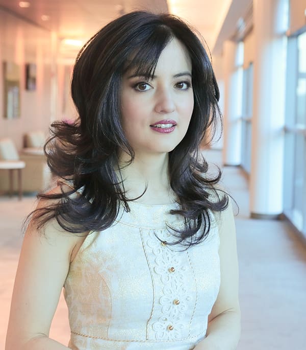
Director of Breast Imaging: Mia Kazanjian, MD
Certified by the American Board of radiology in 2016, Dr. Kazanjian graduated from Brown University, selected to the Phi Beta Kappa Honor Society her junior year. She was accepted early to the Mount Sinai School of Medicine's Humanities and Medicine program, from which she received her medical degree in 2010. Dr. Kazanjian completed her first year of radiology training at New York Presbyterian Hospital, Weill Cornell, with rotations at the Memorial Sloan Kettering Cancer Center. She then spent four years in Palo Alto, California, at Stanford University Hospital, graduating from their radiology residency in 2015. She dedicated her last year of residency at Stanford to fellowship-level training in Body Imaging, focused on advanced modalities of CT, ultrasound, and MRI. She then completed a full year fellowship in Breast Imaging at Stanford University Hospital in 2016.
Dr. Kazanjian has been the Director of Breast Imaging in her practice since 2018. Dr. Kazanjian is also the Co-Director of the Breast Center at Stamford Health.
-
What can I expect during a mammogram?You will be asked to undress from the waist up and change into a gown. A specially trained female technologist will then position you for the exam.
In a typical screening mammogram, each breast is examined separately, with two views of each breast:
• From above (a cranio-caudal or CC view); and
• From the side (a medio-lateral oblique or MLO view)
During a mammogram, the breast is compressed between two plastic plates, which may cause temporary discomfort in some patients. The compression lasts no more than a few seconds and does not harm the breast. Compression is necessary for the following reasons:
• Compression makes a mammogram more accurate, by reducing motion, and allowing the x-ray beam to pass more uniformly through the breast.
• Compression makes a mammogram safer, by reducing the amount of radiation required for an accurate interpretation. -
How long will my 3D mammogram take?
A routine screening exam generally takes 15 minutes or less.
-
Is 3D mammography safe?
Yes, but be sure to have your mammogram at a fully accredited facility that uses low dose technology, like we do at NRC. The benefit of an accurate diagnosis far outweighs the risk of this small amount of radiation. Get more information from radiologyinfo.org.
It is possible that you will get a “false positive,” since 5-15% of screening mammograms require more testing, such as special mammogram images or ultrasound. Most of these exams show normal results. If there is an abnormal finding, you may need a biopsy for further diagnosis.
Patients must always let the mammography technologist know if they are pregnant. It is recommended that these patients wait until after pregnancy to have a screening mammogram. An alternate screening such as ultrasound may be recommended.
-
Do I need a referral or prescription?
Although most patients are referred by their physician, you may schedule a mammogram without a referral or prescription. Please be aware that this is subject to the requirements of your insurance carrier or HMO, so check with them first to verify the applicable rules and coverage. Please note you must have a primary care physician or OB/GYN before scheduling a screening mammogram. The results of your exam will be sent to your physician.
For diagnostic breast imaging, ultrasounds, and bone density scans, a referral from a physician is required.
-
Why do you need my old mammograms?
Just as a person’s fingerprints are unique, so too are a person’s breasts. What is normal in one person may be abnormal in another. The best way for the radiologist to know that a mammogram is normal is to confirm stability from year to year.
If you have had mammograms in the past at any other facility, it is recommended that we obtain them on your behalf to ensure they will be available at the time of your appointment. We simply require a signed release. Alternatively, you may bring the films with you to your scheduled exam.
-
When will I know my mammography results?
Once your screening mammogram is complete, a radiologist will carefully evaluate the images and meticulously compare your current mammogram with prior exams in looking for any subtle change. If you mammogram is normal, a written report will be sent to your medical provider within three working days, although the vast majority will be available within 24 hours. You will receive a letter within 10 working days. We will also provide optional appointments for real-time results.
Should you need any additional imaging to clarify a finding on your screening mammogram, we will call you within three business days of your mammogram to schedule a follow-up appointment. At the follow-up appointment, the radiologist will ensure that all necessary studies are performed to develop a conclusion, and we will discuss the findings with you before you leave. The results will also be given to you in writing, and your physician will receive a written report.
Diagnostic mammogram results will be available during your appointment and your physician will receive the written report. If a biopsy is recommended, a radiologist will consult with you and you will speak with one of our breast biopsy schedulers to make an appointment before leaving the facility.
-
Why would my radiologist order a follow-up mammogram in just six months, rather than in a year?
If you have a condition that appears benign, our radiologists may recommend a six-month follow-up examination to more closely monitor the condition and to confirm stability.
-
Does a mammogram find all breast cancer?
Mammography is the best test available, but it is not perfect. Between 10-15% of breast cancers may not show up on a mammogram (but 85-90% do!). This makes breast self-examination very important. If you have any questions, do not hesitate to ask us, or ask your doctor. The best way to detect breast cancer early is with combination of tests: your self-examination, your doctor’s examination, and annual screening mammography.
-
What does "dense breast tissue" mean?"
Some people have what’s called "dense breast tissue." In fact, about half of all women over age 40 have dense breast tissue. In general, if your breasts have a lot of dense glandular and connective tissue and not much fatty tissue, your breasts may be considered to be dense. A person's breast density can change through their life.
For a definitive evaluation of breast tissue, you need to have a mammogram. There are four types of breast density, from most to least dense.
• Extremely dense
• Heterogeneously dense
• Scattered fibroglandular density
• Almost entirely fatty breast tissue
Dense breasts (extremely dense and heterogeneously dense) may benefit from additional screening breast ultrasound or MRI.
-
What are breast microcalcifications?
Microcalcifications are tiny deposits of calcium in the breast, usually of varying shape, size, and location. Although breast calcifications are usually benign, changes in the pattern, or new calcifications may indicate the presence of a small or developing breast cancer, or even a pre-cancerous condition. If calcifications are stable from year to year, or clearly benign, no biopsy is necessary. In other cases, a radiologist may recommend a biopsy to determine the cause of the calcifications.
-
What is a solid lump or nodule?
A solid lump or nodule refers to a mass within the breast that contains solid tissue. A lump or nodule could represent a rounded clump of normal tissue, a fibroadenoma (a benign tumor), or possibly a malignancy. These conditions often require further evaluation, such as ultrasound or biopsy.
-
What if I find a lump while pregnant?
If you find a lump or a mass, do not ignore it. See your physician. Often an ultrasound (which uses harmless sound waves, rather than x-rays) can be performed for initial evaluation. If a mammogram is necessary, it is usually safe for the fetus, especially after the 14th week of pregnancy, with the abdomen shielded by a lead apron.
-
Can I have a mammogram while I am nursing?
If you have a breast lump or other problem, it is recommended you have a mammogram or an ultrasound during lactation/nursing to evaluate the problem. Otherwise, if you are just scheduled for a routine or screening examination, it is best to wait approximately six months after terminating nursing. This allows the changes in the breast from pregnancy and nursing to resolve.
-
What if I have breast implants?
According to the latest literature, breast implants neither increase nor decrease a person's risk of breast cancer. Patients with implants should obtain a mammogram, according to the same recommendations as those without breast augmentation. Patients who have implants do require specialized views, called Eklund or displacement views, where the implants are gently pushed back to visualize as much breast tissue as possible. The risk of rupturing the implant is minimal.
-
When and why is an ultrasound used?
Breast ultrasound uses sound waves, rather than x-rays, to image the breast. If a mammogram film shows a mass, or if there is a palpable lump on the breast, this study is performed to determine if the mass is cystic or solid. Sometimes, an ultrasound can help suggest whether or not a mass is suspicious.
Ultrasound does not use radiation. It is often the first breast-imaging test in patients under the age of 35 with a palpable finding. Ultrasound may also be helpful in assessing breast pain.
Ultrasound does not replace mammography, but can be used in conjunction with mammography to obtain additional diagnostic information.
-
When and why is a breast biopsy used?
A breast biopsy means removing breast tissue for examination under the microscope. It is the only definitive way to diagnose the nature of a breast mass/lump or calcification. Approximately 80% of breast biopsies are benign.
A biopsy may require surgery (excisional biopsy), however, most breast biopsies can be accurately performed by placing a specially designed needle into the suspicious area (stereotactic core needle biopsy).
There are several ways to perform a breast biopsy. If you need a breast biopsy, we can help you find a breast specialist who can advise you on which method is the best for your individual condition.
-
How should I prepare for my mammogram?
No specific preparation is required. Please do not use powders, talc, lotion or deodorant on the breast and in the underarm area. For your comfort and convenience, two piece outfits are recommended.
If you have had mammograms in the past at any other facility, you will be asked to sign a release to have us request your prior exams on your behalf, or you can bring the films with you to your scheduled exam.


